Choosing In-Vitro Models for Peptide Research: Cell Lines, Primary Cells and Organoids
A practical comparison of immortalised lines, primary cells, stem-cell-derived cultures, co-cultures and organoids for peptide research, with the quality checks every model needs.
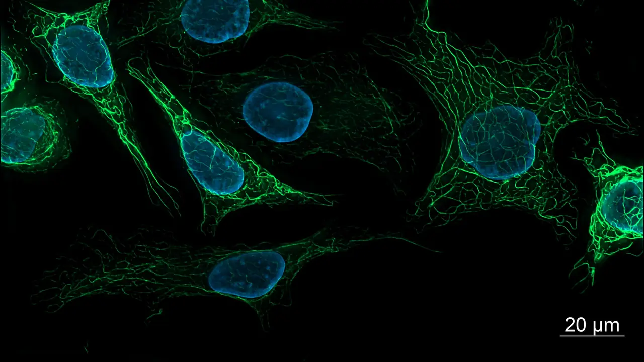
A peptide’s effect is only as meaningful as the model it was measured in. The same compound can look potent in one cell system and inert in another because the receptor, the pathway or the uptake route is missing. Choosing the model is the most important design decision in in-vitro peptide work.
The main options
| Model | Strengths | Limitations |
|---|---|---|
| Immortalised cell lines | Cheap, reproducible, scalable | May lack native receptors; genetic drift with passage |
| Recombinant receptor lines | Clean single-receptor pharmacology | Artificial expression levels |
| Primary cells | Closer to native biology | Donor variability, limited lifespan |
| Stem-cell-derived cells | Human cell types otherwise hard to obtain | Differentiation variability, cost |
| Co-cultures | Cell–cell signalling | More variables to control |
| Organoids and 3D cultures | Tissue-like structure and gradients | Complex readouts, slower |
Match the model to the mechanism
- Receptor pharmacology (for example incretin or GHRH receptors) suits recombinant lines with one receptor each, read out by cAMP or calcium.
- Cell migration and matrix biology suit fibroblasts, endothelial cells and keratinocytes in scratch, Transwell or tube-formation assays.
- Immune signalling needs primary immune cells or well-characterised lines, with rigorous endotoxin control (endotoxin testing).
- Mitochondrial work suits cells with high oxidative capacity, studied by extracellular flux and membrane-potential imaging (MOTS-c and SS-31).
- Neuronal signalling suits primary neurons, differentiated lines or neurospheres (Semax and Selank).
Quality checks every model needs
- Authentication. Confirm human lines by STR profiling, and source from a recognised cell bank.
- Mycoplasma testing. Routine PCR or luminescence testing; mycoplasma changes almost every readout.
- Passage limits. Set a maximum passage number and record it for every experiment.
- Receptor confirmation. Check that the target receptor or pathway is actually expressed (qPCR, western blot or a known agonist response).
- Serum and medium records. Note serum lot and supplement concentrations; peptides can bind serum proteins.
Peptide-specific considerations
- Stability in medium. Proteases in serum and secreted by cells can degrade peptides during long incubations. Consider measuring peptide remaining by LC-MS at the end of the incubation.
- Adsorption. Low-concentration peptides stick to plastics; use low-binding plates or a carrier protein.
- Uptake. Intracellular targets need a cell-permeable peptide or a delivery strategy.
For setting up concentrations and controls across several compounds, see designing multi-compound experiments.
For in-vitro laboratory research only. Not for human or veterinary use. This article summarises laboratory practice and published research. Nothing here is advice about use in people or animals.